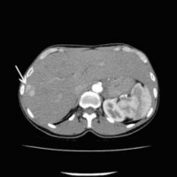
TACE stands for "Transarterial Chemo Embolization" and is a minimally invasive, angiographic form of treatment for malignant liver tumors. Chemoembolization is intended to occlude vessels supplying the tumor and simultaneously apply a chemotherapeutic agent locally.
Transarterial Chemoembolization (TACE)
The treatment can be carried out:
- to prolong life expectancy through local tumor control
- to bridge the time until a possibly planned liver transplantation (bridge-to-transplantation)
- as an additional measure to other tumor-destroying therapies (e.g. radiotherapy, chemotherapy)
- for liver tumors or daughter tumors of other tumors (metastases) in the liver that cannot be operated on and often respond inadequately to anticancer therapy administered systemically (distributed via the bloodstream)
- Before surgery for liver tumors to reduce tumor size.
In TACE, small spherical gelatin or plastic particles containing cell growth inhibiting substances (chemotherapeutic agents) are introduced into the tumor tissue of the liver. This is done via a small catheter (tube) that has previously been inserted into the hepatic artery from the groin. As far as possible, healthy liver tissue is spared so that it can be preserved and remain functional. Chemoembolization has the advantage that the cell growth inhibiting substances can continue to exert their effect locally long after the intervention. Only small amounts of the high-dose chemotherapeutic agents enter the bloodstream.
The intervention itself is initially preceded by an examination of the indication on the basis of all available images and therapy histories. This usually takes place after interdisciplinary consultation with the treating oncologists, surgeons, and radiotherapists within the framework of an interdisciplinary tumor board. If it turns out that TACE is indeed a promising option, the actual treatment takes place during an inpatient stay on our ward. Under local anesthesia (regional anesthesia), an artery is punctured, usually in the groin, with a thin hollow needle. Under X-ray control, a plastic tube (catheter) is selectively inserted into the desired location in the liver. Then the therapeutic substance is injected slowly and in portions (approximately over 30 to 60 minutes) via the positioned catheter into the hepatic artery, and while checking the respective flow conditions. After administration of the substances, the catheter is removed. Afterwards, the patient should be on bed rest for about 24 hours to prevent any secondary bleeding from the groin. The next day, the treatment result is checked by means of computer tomography with, if necessary, planning of a second therapy session after approx. 2 - 4 weeks. Discharge depends on the clinical condition, usually about 2-3 days after therapy.
The treatment is usually well tolerated. However, some patients experience upper abdominal pain, nausea and fever for a short time (within the first few days, possibly already during the treatment), in the sense of a so-called post embolisation syndrome. This can usually be treated very well with medication and usually subsides after about 1 week. Very rarely, serious side effects can occur, e.g. if, despite all precautionary measures, the embolisation material is carried from one vessel into a neighbouring one.
We make sure that the patient can lie comfortably and quietly for the entire duration of the treatment. During the treatment, the patient's vital parameters (pulse, blood pressure) are checked at regular intervals and also documented. Each patient is given venous access so that infusions or medication can be administered during the procedure. A local anaesthetic is injected into the skin and subcutaneous fatty tissue in the area of the access route in the groin. With this medication, the procedure is usually painless. Occasionally, post embolisation syndrome occurs after the procedure. Minutes to hours after the procedure, there may be more or less severe upper abdominal pain. Nausea, vomiting and fever are also observed. However, these symptoms can be controlled very well with medication. In rare cases, these symptoms can last up to a week after the therapy.
In general, embolisation is successful when the tissue to be embolised is supplied by only one artery. Embolisation becomes more difficult when several vessels need to be probed or occluded. Occasionally, several treatments are necessary until complete success is achieved.
Before discharge, a CT scan of the upper abdomen is performed to check the success of the treatment. A follow-up is carried out with CT and/or MRI and blood checks at 3-month intervals. The patient automatically receives an appointment for this (if desired) via our outpatient department.
 Computertomographische Darstellung eines hepatozellulären Karzinoms (Pfeil)
(Bild 1 von 5)
Vorwärts »
 « Zurück
Computertomographische Dokumentation der Embolisateinlagerung innerhalb des Tumors nach transarterieller Chemoembolisation (TACE)
(Bild 2 von 5)
Vorwärts »
 « Zurück
Superselektive transarterielle Chemoembolisation (TACE) mit Sondierung eines tumorversorgenden Gefäßes 1
(Bild 3 von 5)
Vorwärts »
 « Zurück
Superselektive transarterielle Chemoembolisation (TACE) mit Sondierung eines tumorversorgenden Gefäßes 2
(Bild 4 von 5)
Vorwärts »
 « Zurück
Superselektive transarterielle Chemoembolisation (TACE) mit Sondierung eines tumorversorgenden Gefäßes 3
(Bild 5 von 5)


