Thermoablation (MWA/RFA/Laser therapy)
The treatment is divided into the following steps, illustrated using the liver and lungs as examples:
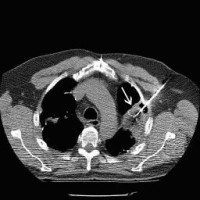
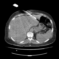
After taking preparatory images and determining the access route, the skin is disinfected and the puncture site is sterilely covered. Under local anesthesia of the puncture site and administration of analgesic substances, the tumor is punctured and the probe is placed in the target volume under sonographic or CT/MRI control.
Now the lesion is heated by microwave ablation, radiofrequency current, or laser, depending on the therapy method, and destroyed/cooked in intervals of 5 to 30 min. Necrosis is produced. The destroyed tissue is degraded by the body's own cells over the next few months.
The patient is then monitored for a few more hours and transferred back to the ward. If there are no complications, the patient can be discharged on the 2nd day after the procedure.
The success of the therapy is monitored by CT and/or MRI and blood tests at 3-month intervals. For this purpose, the patient (if desired) automatically receives an appointment through our outpatient clinic.
 ...
...
Images: CT-guided radiofrequency ablation of a lung metastasis (arrow, left), CT-guided microwave ablation of a liver metastasis (right).


